ANTI BIOTIN MONOCLONAL ANTIBODY from Mouse hybridoma
| Appearance: | Solution with PBS buffer containing 0.05% NaN₃ |
|---|---|
| Grade: | GradeⅡ |
| Immunogen: | Biotin-KLH |
| Purity: | ≥ 90%(GPC) |
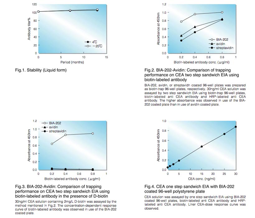
| Stability: | Stable at 4°C and -20°C for at least 12 months(Fig.1) |
|---|---|
| Isotype: | IgG₁ |
BIA-202
APPLICATIONS
This monoclonal antibody is useful as a trap reagent for biotin-labeled material such as antibodies, DNA probes and allergens. (Fig.2, 3, 4)
Examples
Examples in detail
Fig.2. BIA-202-Avidin: Comparison of trapping performance on CEA two step sandwich EIA using biotin-labeled antibody
Biotin-trap 96-well plates was prepared as shown below. 50uL of 0.01mg/ml BIA-202, avidin, or streptavidn in 0.1M phosphate buffer pH7.0 were added to 96-well polystyrene plate and incubated at 25°C for 2 hr, respectively. After discarding these solution, 300uL of blocking buffer was added to the plates and incubated at 25°C for 1 hr.
After washing PBS-T, 25ul of 30ng/ml CEA solution and 25ul of 0.2, 0.4, 0.8ug/ml biotin-labeled anti-CEA monoclonal antibody were added and incubated at 37°C for 1hr, respectively. After washing PBS-T, 50ul of HRP- labeled anti-CEA monoclonal antibody were added to the plate and incubated at 37°C for 1 hr. After washing PBS-T, 50ul of TMB solution was added to the plate and incubated at room temperature for 10 min. After 50ul of 1N sulfuric acid solution was added to the plate, absorbance of each well was measured at 450nm.
The higher absorbance was observed in use of the BIA-202 coated plate than in use of avidin coated plate.
Fig.3. BIA-202-Avidin: Comparison of trapping performance on CEA two step sandwich EIA using biotin-labeled antibody in the presence of D-biotin
30ng/ml CEA solution containing 2mg/L D-biotin was assayed by the method mentioned in Fig.2. The concentration-dependent response curve of biotin-labeled antibody was observed in use of the BIA-202 coated plate.
Fig.4. One step sandwich EIA with BIA-202 coated 96-well polystyrene plate
Biotin-trap 96-well plates were prepared as shown Fig.2. After washing PBS-T, 25ul of the standard solution (hCEA) and 25ul of pre-mixed conjugate solution (containing 0.4ug/ml biotin-labeled anti-CEA monoclonal antibody and HRP-labeled anti-CEA monoclonal antibody) were added to the plate and incubated at 37°C for 1 hr. After washing PBS-T, the enzyme reaction and the measurement were done as shown Fig.2.
Liner CEA-dose response curve was observed.
To get a quote, contact us at info@toyobousa.com, or INQUIRY.