LEUCINE DEHYDROGENASE from Bacillus sp. ¹⁾
LED-201
| Appearance: | White amorphous powder, lyophilized | ||
|---|---|---|---|
| Activity: | GradeⅡ 20U/mg-solid or more (containing approx. 70% of stabilizers) |
||
| Contaminants: | Leucylpeptide decomposing enzymes (Leu-Val) ≤1.0×10⁻²% (Leu-Gly-Gly) ≤1.0×10⁻²% NADH oxidase ≤1.0×10⁻²% |
||
| Stabilizers: | 2-Mercaptoethanol, L-cysteine, dithiothreitol, ethylenediaminetetraacetate | ||
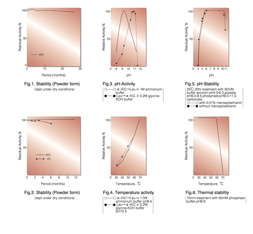
| Stability: | Stable at -20°C for at least One year (Fig.1) |
|---|---|
| Molecular weight ² ⁾: | 245,000 |
| Michaelis constants ² ⁾: | 1.0×10⁻³M (L-Leucine), 3.9×10⁻⁴M (NAD⁺), 3.5×10⁻⁵M (NADH), 3.1×10⁻⁴M [α-Ketoisocaproate (α-KIC)], 2.0×10⁻¹M (NH₃) |
| Structure ² ⁾: | 6 subunits per enzyme molecule |
| Inhibitors ² ⁾: | Na₂S, Hg⁺⁺, Cu⁺⁺, Co⁺⁺, Mg⁺⁺, p-chloromercuribenzoate |
| Optimum pH : | 10.5-10.8 (L-Leu→α-KIC), 9.4 (α-KIC→L-Leu )(Fig.3) |
| Optimum temperature : | above 70°C(Fig.4) |
| pH Stability : | pH 5.5-10.5 (25°C, 20hr)(Fig.5) |
| Thermal stability : | below 60°C (pH 6.9, 10min)(Fig.6) |
| Substrate specificity: | (Table 1) |
APPLICATIONS ⁴⁾
This enzyme is useful for enzyme determination of L-leucine and the activity of leucine amino- peptidase.
ASSAY
Principle:
leucine dehydrogenase
L-Leucine+NAD⁺+H₂O ►α-Ketoisocaproate+NADH+NH₃+H⁺
The appearance of NADH is measured at 340nm by spectrophotometry.
Unit definition:
One unit causes the formation of one micromole of NADH per minute under the conditions described below.
Method:
| A. L-Leucine solution: | 20mM L-leucine in 0.2M glycine-KCl-KOH buffer, pH 10.5 (Prepare freshly) |
|---|---|
| B. NAD⁺ solution: | 12.5mM (Should be prepared fresh) |
| C. Enzyme diluent: | 25mM K-phosphate buffer, pH 7.2 |
Procedure
| Concentration in assay mixture | |
|---|---|
| Glycine buffer | 0.18 M |
| L-Leucine | 18m M |
| NAD⁺ | 1.1mM |
1. Prepare the following reaction mixture in a cuvette (d=1.0cm) and equilibrate at 37°C for about 5 minutes.
3.0ml Substrate solution (A)
0.3ml NAD⁺ solution (B)
2. Add 0.05ml of the enzyme solution* and mix by gentle inversion.
3. Record the increase in optical density at 340nm against water for 2 to 3 minutes in a spectrophotometer
thermostated at 37°C, and calculate the ΔOD per minute from the initial linear portion of the curve (ΔOD test).
At the same time, measure the blank rate (ΔOD blank) by using the same method as the test except that the enzyme diluent (C) is added instead of the enzyme solution.
* Dissolve the enzyme preparation in ice-cold enzyme diluent (C) (ca. 5mg/ml) and dilute to 0.25−0.33U/ml with the same buffer, immediately before assay.
Calculation
Activity can be calculated by using the following formula :
ΔOD/min (ΔOD test-ΔOD blank)×Vt×df
Volume activity (U/ml) = = ΔOD/min×10.77×df
6.22×1.0×Vs
Weight activity (U/mg)=(U/ml)×1/C
- Vt
- : Total volume (3.35ml)
- Vs
- : Sample volume (0.05ml)
- 6.22
- : Millimolar extinction coefficient of NADH (㎠/micromole)
- 1.0
- : Light path length (cm)
- df
- : Dilution factor
- C
- : Enzyme concentration in dissolution (c mg/ml)
REFERENCES
- K.Soda et al.; Biochem.Biophys.Res.Commun., 44, 931 (1971).
- T.Ohshima et al.; J.Biol.Chem., 253, 5719 (1978).
| Substrate (10mM) | Relative activity(%) | Substrate (10mM) | Relative activity(%) |
|---|---|---|---|
| L-Leucine | 100 | α-Ketoisocaproate | 100 |
| L-Valine | 74 | α-Ketoisovalerate | 126 |
| L-Isoleucine | 58 | α-Ketovalerate | 76 |
| L-Norvaline | 41 | α-Ketobutyrate | 57 |
| L-Norleucine | 10 | α-Ketocaproate | 46 |
| L-Methionine | 0.6 | Inert: Pyruvate, α-Ketoglutarate, Phenylpyruvate, Oxaloacetate, Glyoxylate |
|
| L-Cysteine | 0.3 | ||
| Inert: L-Ala, L-Glu, L-Thr, L-Ser Gly, L-Phe, L-Lys, L-Arg, D-Leu, D-Val, D-Ile |
|||
To get a quote, contact us at info@toyobousa.com, or INQUIRY.