L-α-GLYCEROPHOSPHATE OXIDASE from Microorganism, G3O-311
| Appearance: | Yellowish amorphous powder, lyophilized | ||
|---|---|---|---|
| Activity: | GradeⅢ 15 U/mg-solid or more (containing approx. 60% of stabilizers) |
||
| Contaminants: | Lactate oxidase ≤2.0×10⁻⁴%| Phosphatase ≤1.0×10⁻³%| |
||
| Stabilizers: | Sucrose, FAD | ||
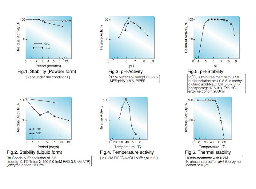
| Stability: | Stable at -20°C for at least 6 months(Fig.1) |
|---|---|
| Molecular weight : | approx. 93,000 (by gel filtration) |
| Isoelectric point: | 4.6±0.1 |
| Michaelis constant : | 2.3×10⁻³M (L-α-Glycerophosphate) |
| Inhibitors: | SH-reagents, ionic detergents, metal ions, etc. |
| Optimum pH : | 6.5-7.0(Fig.3) |
| Optimum temperature: | 40°C(Fig.4) |
| pH Stability: | 5.0-7.5 (25°C, 60min)(Fig.5) |
| Thermal stability: | below 45°C (pH 6.5, 10min)(Fig.6) |
| Effect of various chemicals: | (Table 1) |
APPLICATIONS
This enzyme is useful for enzymatic determination of triglyceride when coupled with lipoprotein lipase (LPL-311, LPL-314) and glycerokinase (GYK-301, GYK-311) in clinical analysis.
G3O-311
ASSAY
Principle:
L-α-glycerophosphate oxidase
L-α-Glycerophosphate+O₂ ► Dihydroxyacetonephosphate+H₂O₂
peroxidase
2H₂O₂+4-Aminoantipyrine+EHSPT ► Quinoneimine dye+4H₂O
The appearance of quinoneimine dye is measured at 555nm by spectrophotometry.
Unit definition:
One unit causes the formation of one micromole of hydrogen peroxide (half a micromole of quinoneimine dye) per
minute under the conditions described below.
Method:
| A. D,L-α-Glycerophosphate: solution | 1.5M [Weigh 48.63g of D,L-α-Glycerophosphate (disodium salt, MW= 324.17), dissolve in 60ml of H₂O and after adjusting the pH to 6.5±0.05 at 25°C with 4.0 N HCl, fill up to 100ml with H₂O] (Stable for two weeks if stored at 0-4°C) |
|---|---|
| B. PIPES-NaOH buffer, pH 6.5: | 0.5M [Weigh 15.12g of PIPES (MW=302.36), suspend in 60ml of H₂O, dissolve with 10 N NaOH. After adjusting the pH to 6.5±0.05 at 25°C with 10 N NaOH, fill up to 100ml with H₂O] (Stable for two weeks if stored at 0- 4°C) |
| C. 4-AA solution: | 28mM [569mg 4-aminoantipyrine (MW=203.25)/100ml of H₂O](Stable for one week if stored at 4°C in a brownish bottle) |
| D. EHSPT (TOOS) solution: | 20mM [591mg N-ethyl-N-(2-hydroxy-3-sulfopropyl)-m-toluidine (MW= 295.3)/100ml of H₂O] (Stable for one week if stored at 4°C in a brownish bottle) |
| E. Peroxidase solution: | 0.05% [50mg peroxidase (110 purpurogallin units/mg)/100ml of H₂O] (Should be prepared fresh) |
| F. Enzyme diluent: | 20mM PIPES-NaOH buffer, pH 6.5 contg. 0.5M NaCl |
Procedure
| Concentration in assay mixture | |
|---|---|
| PIPES-NaOH buffer | 193 mM |
| NaCl | 19.2 mM |
| D,L-α-Glycerophosphate | 577 mM |
| 4-Aminoantipyrine | 1.3 mM |
| EHSPT | 0.96mM |
| Peroxidase | ca.5.3 U/ml |
1. Prepare the following working solution (40 tests) in a brownish bottle and store on ice.
40 ml Substrate solution (A)
40 ml PIPES-NaOH buffer, pH 6.5 (B)
5 ml 4-AA solution (C)
5 ml EHSPT solution (D)
10 ml Peroxidase solution (E)
2. Pipette 2.5ml of working solution into a cuvette (d=1.0cm) and equilibrate at 30°C for about 5 minutes.
3. Add 0.10 ml of the enzyme solution* and mix by gentle inversion.
4. Record the increase in optical density at 555nm against water for 3 to 4 minutes in a spectrophotometer
thermostated at 30°C, and calculate theΔOD per minute from the initial linear portion of the curve (ΔOD test).
At the same time, measure the blank rate (ΔOD blank) by using the same method as theΔOD test except that the enzyme diluent is added instead of enzyme solution.
* Dissolve the enzyme preparation in ice-cold enzyme diluent (F), dilute to 0.05-0.20U/ml with the same buffer and store on ice.
Calculation
Activity can be calculated by using the following formula :
ΔOD/min (ΔOD test−ΔOD blank ) ×Vt × df
Volume activity (U/ml) = =ΔOD/min×1.739×df
29.9×1/2×1.0×Vs
Weight activity (U/mg)=(U/ml)×1/C
- Vt
- : Total volume (2.60ml)
- Vs
- : Sample volume (0.10ml)
- 29.9
- : Millimolar extinction coefficient of quinoneimine dye under the assay condition (㎠/micromole)
- 1/2
- : Factor based on the fact that one mole of H₂O₂ produces half a mole of quinoneimine dye.
- 1.0
- : Light path length (cm)
- df
- : Dilution factor
- C
- : Enzyme concentration in dissolution (c mg/ml)
REFERENCES
- L-α-Glycerophosphate oxidase from
- C.Gancedo, J.M.Gancedo and A.Sols; European J.Biochem, 5,165 (1968).
- W.S.Kistler, C.A.Hirsch, N.R.Cozzarelli and E.C.C.Lin; J.Bacteriology, 100, 1133 (1969).
- C.F.Strittmatter; J.Biol.Chem., 234, 2794 (1959).
- N.J.Jacobs and P.J.VanDemark; Arch.Biochem. and Biophys., 88, 250 (1960).
- L.K.Koditschek and W.W.Umbreit; J.Bacteriology, 98,1063 (1969).
- T.W.Esders and C.A.Michrina; J.Biol.Chem., 254, 2710 (1979).
Yeast
Escherichia coli
Lactobacillus casei
Streptococcus feacalis
Streptococcus feacium
| Chemical | Concn.(mM) | Residual activity |
Chemical | Concn.(mM) | Residual activity |
|---|---|---|---|---|---|
| None | − | 100% | PCMB | 2.0 | 3.3 |
| Metal salt | 2.0 | MIA | 2.0 | 3.3 | |
| MgCl₂ | 98 |
NaF | 2.0 | 100 | |
| CaCl₂ |
107 | NaN₃ | 200 | 96 |
|
| Ba(OAc)₂ | 99 | EDTA | 100 | 99 | |
| FeCl₂ | 0.3 | o-Phenanthroline | 2.0 | 95 | |
| FeCl₃ | 0.8 | α,α′-Dipyridyl | 1.0 | 99 | |
| CoCl₂ | 103 | Borate | 50 | 10 | |
| MnCl₂ | 100 | IAA | 2.0 | 2.8 |
|
| ZnSO₄ | 99 | NEM | 2.0 | 97 | |
| NiCl₂ | 98 | Hydroxylamine | 2.0 | 9.7 | |
| CuSO₄ | 7.5 | Triton X-100 | 0.10% | 106 | |
| Pb(OAc)₂ | 87 | Brij 35 | 0.10% | 3.9 | |
| AgNO₃ | 79 | Tween 20 |
0.10% | 3.4 | |
| HgCl₂ | 96 | Span 20 | 0.10% | 94 | |
| Na-cholate | 0.10% | 92 | |||
| SDS | 0.05% | 4.8 | |||
| DAC | 0.05% | 70 |
Ac, CH₃CO; PCMB, p-Chloromercuribenzoate; MIA, Monoiodoacetate; EDTA, Ethylenediaminetetraacetate; IAA, Iodoacetamide; NEM, N-Ethylmaleimide; SDS, Sodium dodecyl sulfate; DAC, Dimethyl-benzyl-alkyl-ammonium chloride.
To get a quote, contact us at info@toyobousa.com, or INQUIRY.