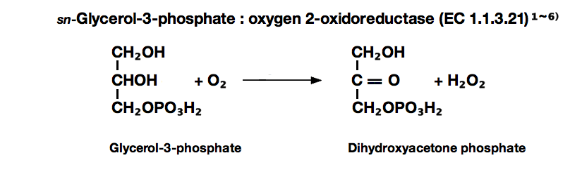
L-α-GLYCEROPHOSPHATE OXIDASE from Pediococcus sp.
| Appearance: | Yellowish amorphous powder, lyophilized | ||
|---|---|---|---|
| Activity: | GradeⅢ 40 U/mg-solid or more (containing approx. 40% of stabilizers) |
||
| Contaminant: | Lactate oxidase ≤1.0×10⁻³% |
||
| Stabilizers: | Sucrose, FAD | ||
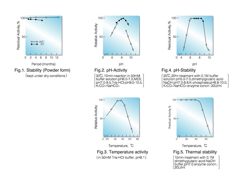
| Stability: | Stable at -20°C for at least one year (A decrease in activity of ca. 10% may occur at 5°C within 6 months.)(Fig.1) |
|---|---|
| Molecular weight : | approx. 76,000 (by gel filtration) |
| Isoelectric point: | 4.1±0.1 |
| Michaelis constants : | 3.2×10⁻³M (L-α-Glycerophosphate), 6.8×10⁻³M (D, L-form) |
| Inhibitors: | Ionic detergents (SDS, LBS, etc.), Hg⁺⁺, Ag⁺ |
| Optimum pH : | 8.0-8.5(Fig.2) |
| Optimum temperature: | 35-40°C(Fig.3) |
| pH Stability: | pH 6.5-8.5 (25°C, 20hr)(Fig.4) |
| Thermal stability: | below 40°C (pH 7.0, 15min)(Fig.5) |
| Substrate specificity: | The enzyme has the highest specificity for L-form of α-glycerophosphate. |
| Effect of various chemicals: | (Table 1) |
APPLICATIONS
This enzyme is useful for enzymatic determination of triglyceride when coupled with lipoprotein lipase (LPL-311, LPL-314) and glycerokinase (GYK-301, GYK-311) in clinical analysis.
G3O-301
ASSAY
Principle:
L-α-glycerophosphate oxidase
L-α-Glycerophosphate+O₂ ► Dihydroxyacetonephosphate+H₂O₂
peroxidase
2H₂O₂+4-Aminoantipyrine+Phenol ► Quinoneimine dye+4H₂O
The appearance of quinoneimine dye is measured at 500nm by spectrophotometry.
Unit definition:
One unit causes the formation of one micromole of hydrogen peroxide (half a micromole of quinoneimine dye) per
minute under the conditions described below.
Method:
| A. D,L-α-Glycerophosphate: solution | 0.2M [Weigh 6.48g of D, L-α-glycerophosphate (disodium salt, MW=324.17), dissolve in 80ml of 0.125M Tris-HCl buffer, pH 8.0 contg. 0.125% of Triton X-100 and after adjusting the pH to 8.1 at 25°C with 2.0N HCl, fill up to 100ml with H₂O.] (Stable for two weeks if stored at 0-5°C) |
|---|---|
| B. 4-AA solution: | 0.1% (100mg of 4-aminoantipyrine /100ml of H₂O)(Store at 4°C in a brownish bottle) |
| C. Phenol solution: | 0.1% (100mg phenol/100ml of H₂O)(Store at 4°C in a brownish bottle) |
| D. Peroxidase solution: | 0.025%[25mg peroxidase (110 purpurogallin units/mg)/100ml of H₂O] (Should be prepared fresh) |
| E. SDS solution: | 0.25% (1.25g sodium dodecyl sulfate/500ml of H₂O) |
| F. Enzyme diluent: | 20mM Tris-HCl buffer, pH 7.5 contg. 0.2% of BSA |
Procedure
| Concentration in assay mixture | |
|---|---|
| Tris-HCl buffer | 50 mM |
| Glycerophosphate | 98 mM |
| 4-Aminoantipyrine | 0.48mM |
| Phenol |
2.1 mM |
| Triton X-100 | 0.049 % |
| POD | ca.5.4 U/ml |
1. Prepare the following working solution (100 tests) in a brownish bottle and store on ice.
50 ml Substrate solution (A)
10 ml 4-AA solution (B)
20 ml Phenol solution (C)
20 ml Peroxidase solution (D)
2. Pipette 1.0ml of working solution into a test tube and equilibrate at 37°C for about 5 minutes.
3. Add 0.02ml of the enzyme solution* and mix.
4. After exactly 10 minutes at 37°C, add 2.0ml of SDS solution (E) to stop the reaction and measure the optical
density at 500nm against water (OD test).
At the same time, prepare the blank by first mixing 1.0ml of working solution with 2.0ml of SDS solution
after 10min-incubation at 37°C, followed by addition of the enzyme solution (OD blank).
* Dissolve the enzyme preparation in ice-cold enzyme diluent (1.0mg/ml or more) and dilute to 0.15-0.35U/ml
with the same buffer, immediately before assay.
Calculation
Activity can be calculated by using the following formula :
ΔOD (OD test−OD blank ) ×Vt × df
Volume activity (U/ml) = =ΔOD×2.2707×df
13.3×1/2× t ×1.0×Vs
Weight activity (U/mg)=(U/ml)×1/C
- Vt
- : Total volume (3.02ml)
- Vs
- : Sample volume (0.02ml)
- 13.3
- : Millimolar extinction coefficient of quinoneimine dye under the assay condition (㎠/micromole)
- 1/2
- : Factor based on the fact that one mole of H₂O₂ produces half a mole of quinoneimine dye.
- t
- : Reaction time (10 minutes)
- 1.0
- : Light path length (cm)
- df
- : Dilution factor
- C
- : Enzyme concentration in dissolution (c mg/ml)
REFERENCES
- L-α-Glycerophosphate oxidase from
- C.Gancedo, J.M.Gancedo and A.Sols; European J.Biochem, 5,165 (1968).
- W.S.Kistler, C.A.Hirsch, N.R.Cozzarelli and E.C.C.Lin; J.Bacteriology, 100, 1133 (1969).
- C.F.Strittmatter; J.Biol.Chem., 234, 2794 (1959).
- N.J.Jacobs and P.J.VanDemark; Arch.Biochem. and Biophys., 88, 250 (1960).
- L.K.Koditschek and W.W.Umbreit; J.Bacteriology, 98,1063 (1969).
- T.W.Esders and C.A.Michrina; J.Biol.Chem., 254, 2710 (1979).
Yeast
Escherichia coli
Lactobacillus casei
Streptococcus feacalis
Streptococcus feacium
| Chemical | Concn.(mM) | Residual activity |
Chemical | Concn.(mM) | Residual activity |
|---|---|---|---|---|---|
| None | − | 100% | PCMB | 2.0 | 100 |
| Metal salt | 2.0 | MIA | 2.0 | 93 | |
| MgCl₂ | 100 |
NaF | 2.0 | 101 | |
| CaCl₂ |
100 | NaN₃ | 20 | 95 |
|
| Ba(OAc)₂ | 95 | EDTA | 5.0 | 105 | |
| FeCl₃ | 98 | o-Phenanthroline | 2.0 | 97 | |
| CoCl₂ | 96 | α,α′-Dipyridyl | 2.0 | 95 | |
| MnCl₂ | 100 | Borate | 50 | 92 |
|
| Zn(OAc)₂ | 100 | Triton X-100 | 0.10% | 105 | |
| NiCl₂ | 99 | Brij 35 | 0.10% | 103 | |
| CuSO₄ | 100 | SDS | 0.05% | 1.5 | |
| Pb(OAc)₂ | 97 | LBS | 0.05% | 1.4 | |
| AgNO₃ | 24 | Na-cholate |
0.10% | 106 | |
| HgCl₂ | 34 |
Ac, CH₃CO; PCMB, p-Chloromercuribenzoate; MIA, Monoiodoacetate; EDTA, Ethylenediaminetetraacetate; SDS, Sodium dodecyl sulfate; LBS, Sodium laurylbenzenesulfonate.
To get a quote, contact us at info@toyobousa.com, or INQUIRY.